
Pancreas · Visual journey
How can the pancreas make the eyes turn yellow?
The pancreas sits in the upper abdomen. To understand the connection, first we need to locate it and follow its ducts.
Scroll to look closer.
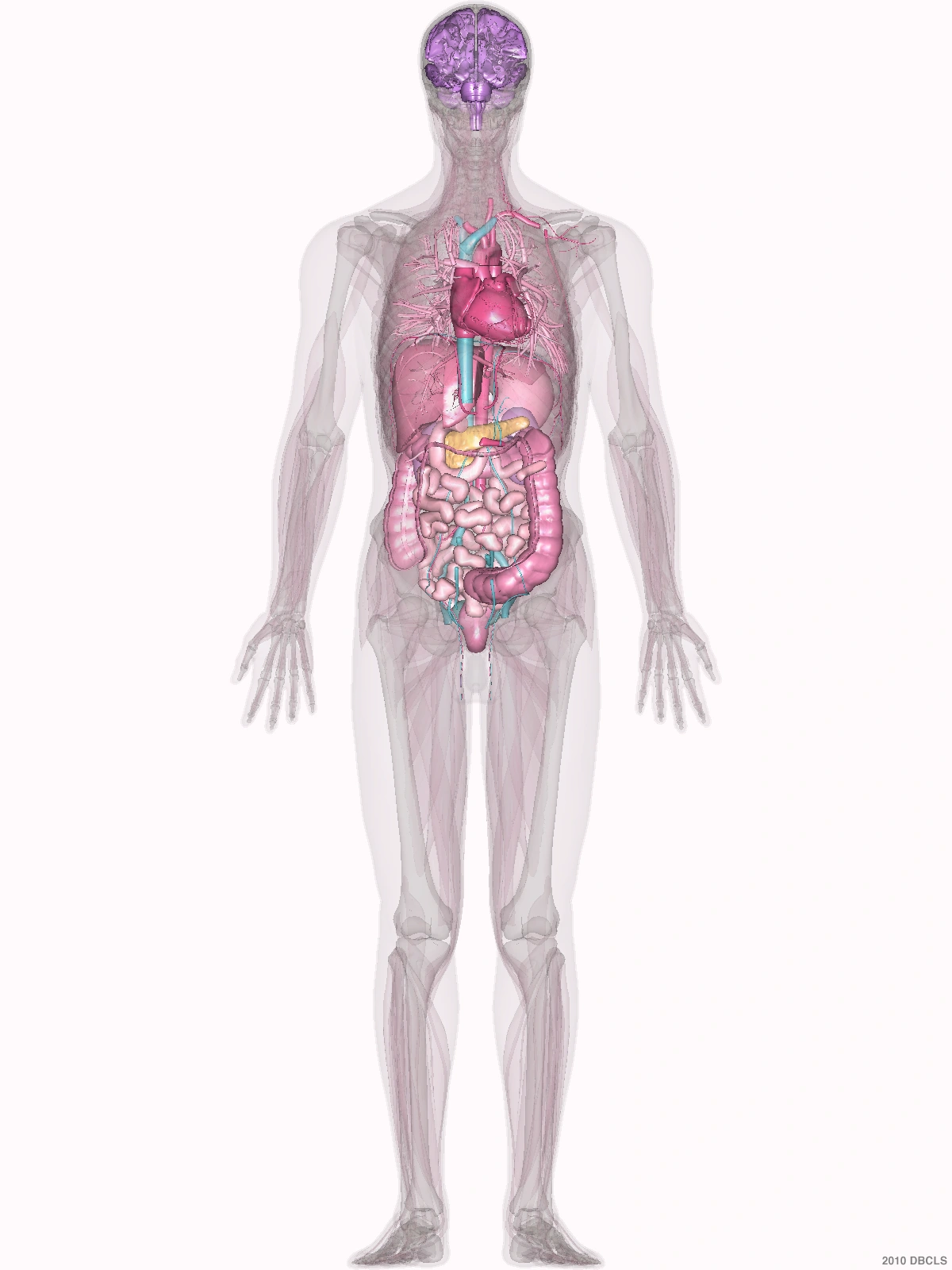
The organ
Behind the stomach.
The head of the pancreas sits beside the duodenum, the first part of the small intestine. Bile passes nearby on its way to the intestine.
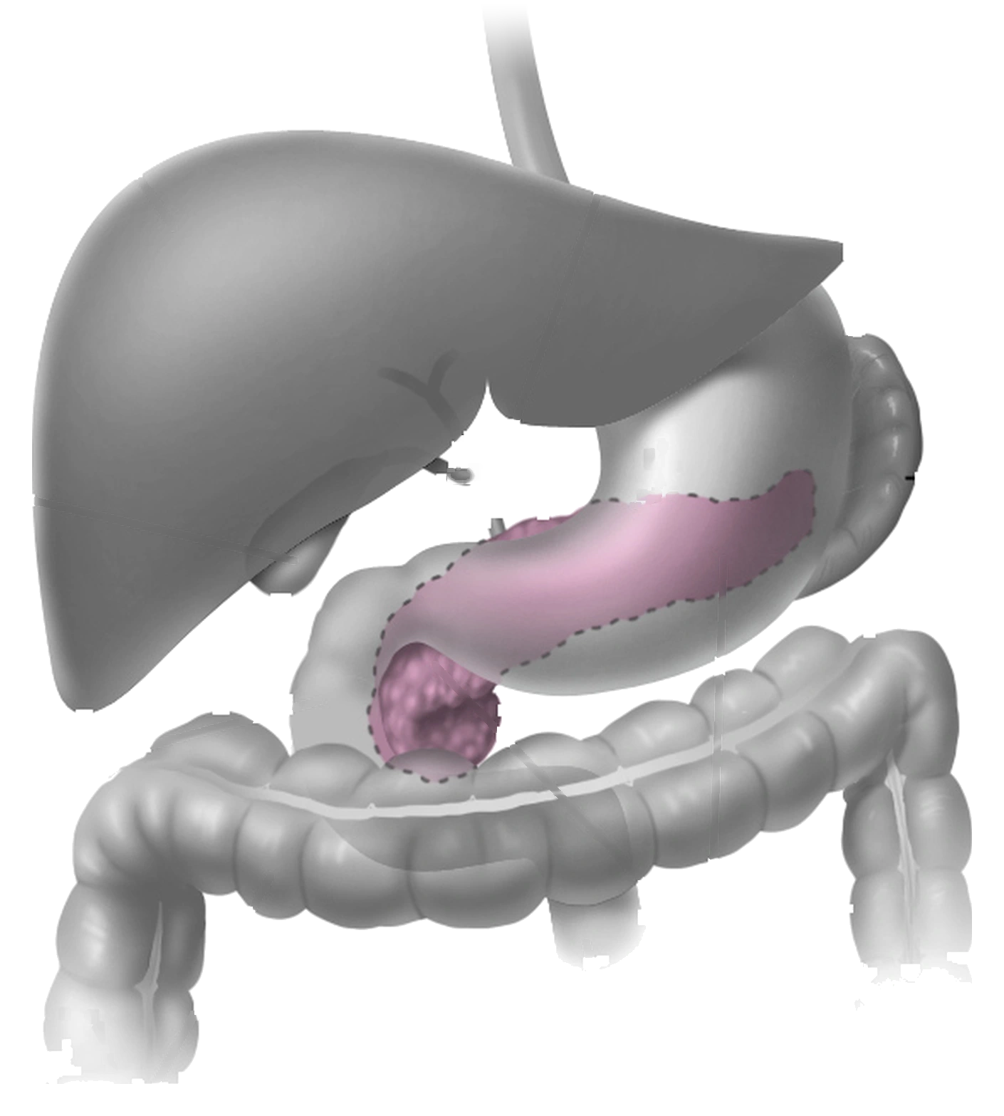 PancreasIt stays hidden behind the stomach, against the spine
On the way to the intestine
Two ducts carry different fluids.
The pancreatic duct carries juices that help digest food. The bile duct brings bile from the liver. Both reach the duodenum: notice how they come together beside the head of the pancreas.
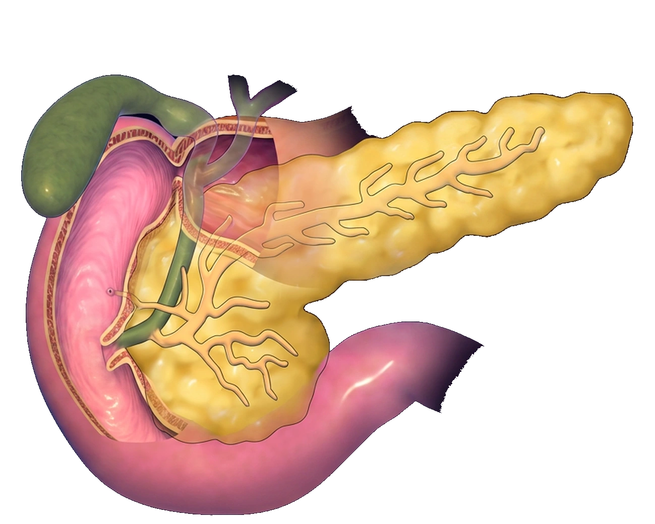 Bile ductCarries bile from the liverPancreatic ductCarries digestive juices
From the duct to its cells
Cells line the pancreatic duct.
Here are normal cells from that lining, collected in a pancreatic sample. Their nuclei, the dark areas inside each cell, are regularly sized and spaced.
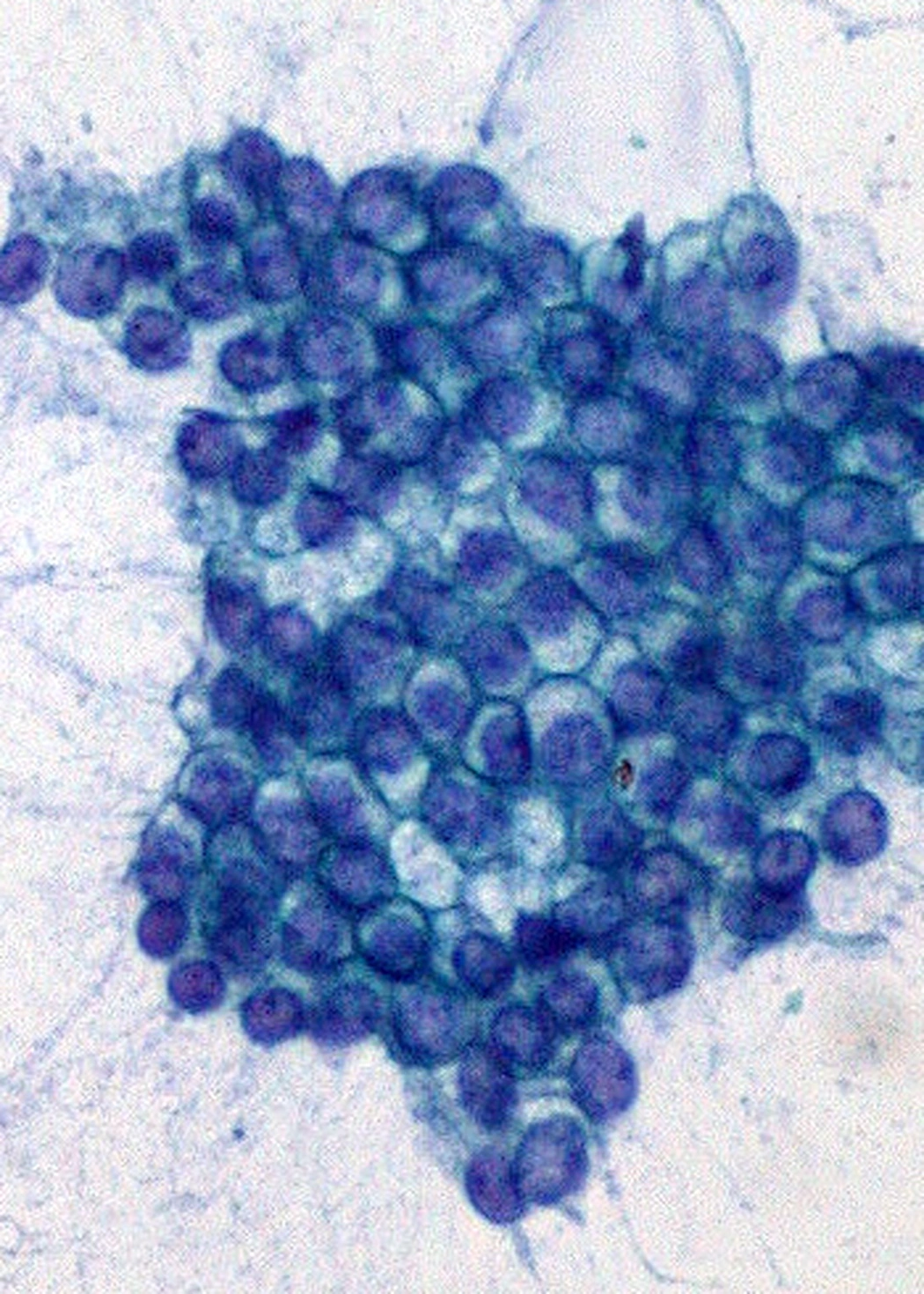
A neighboring duct
A mass can block the flow of bile.
Common bile ductIf it becomes obstructed, bile does not drain properlyA mass in the head of the pancreas can press on the bile duct from outside. If bile cannot drain properly, bilirubin, a yellow pigment, builds up and causes jaundice: yellow skin or eyes.
Gallstones and liver disease are more common causes of jaundice. Yellowing alone cannot tell us the cause.
Finding out what the mass is
A sample lets us examine the cells.
When a mass needs to be investigated, a sample lets doctors examine its cells. Compare the normal pancreatic duct lining you just saw with pancreatic adenocarcinoma, a cancer of glandular cells.
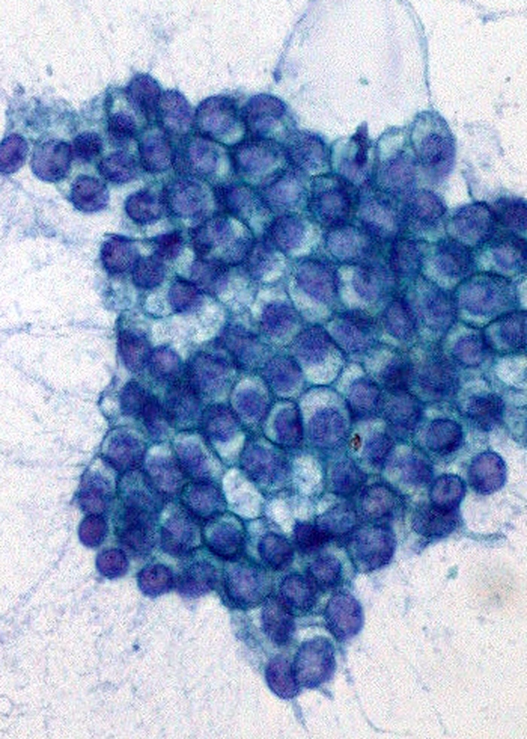 Like a honeycomb
The nuclei, the dark areas inside the cells, are small and regularly spaced.
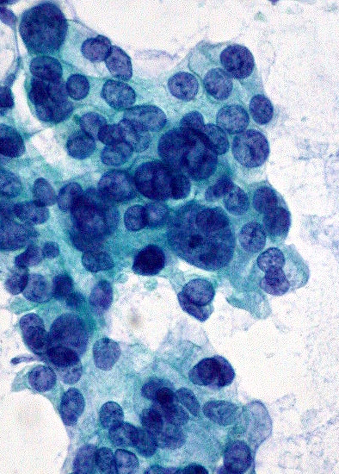 No even spacing
In this cancer, the nuclei are larger, uneven, and overlapping. This is one of the changes assessed by a pathologist, a doctor who examines cells.
What you can do
If your eyes turn yellow, find out why.
If your skin or eyes turn yellow, seek urgent medical care, even without pain. There are several possible causes, and identifying the cause matters.
When seeking care, explain when the color change began. This helps guide the assessment; do not delay seeking care to keep a record.
I’m your friend, Dr. Carlo De la Sancha.
Medical transparencySources and scope
Medical evidence, image provenance, and the editorial transformations applied.
- What symptom information helps during a medical visit .. National Cancer Institute
- Pancreatic juices and bile: their routes to the intestine .. NIDDK
- Bile duct compression, bilirubin and jaundice .. American Cancer Society
- Anatomical body model .. BodyParts3D / Anatomography · DBCLS, CC BY 4.0
- Illustration of the pancreas and nearby organs .. Don Bliss, National Cancer Institute, Public domain
- Illustration of the head of the pancreas and its ducts .. BruceBlaus, CC BY 3.0
- Pancreatic aspirate with adenocarcinoma and normal duct lining in the same field .. Ed Uthman, MD, CC BY 2.0
- Where the pancreas sits and how it is divided .. StatPearls, “Anatomy, Abdomen and Pelvis: Pancreas”, NCBI Bookshelf
- How the common bile duct runs in relation to the head of the pancreas .. StatPearls, “Anatomy, Abdomen and Pelvis: Pancreas”, NCBI Bookshelf
- Bile duct obstruction and painless jaundice .. StatPearls, “Pancreatic Cancer”, NCBI Bookshelf
- The many causes of jaundice .. MedlinePlus, “Jaundice”, U.S. National Library of Medicine
- How a gallstone attack presents .. National Institute of Diabetes and Digestive and Kidney Diseases (NIDDK)
- Endoscopic ultrasound guided aspiration and biopsy in solid masses .. American Society for Gastrointestinal Endoscopy guideline on solid pancreatic masses, Gastrointestinal Endoscopy, 2024 (PMID 39387777)
- What separates benign ductal epithelium from adenocarcinoma in cytology .. Iqbal et al., “Endoscopic-ultrasound-guided fine-needle aspiration and the role of the cytopathologist in solid pancreatic lesion diagnosis”, Pathology Research International, 2012
- Urgent care for jaundice .. NHS, Jaundice
The anatomical illustrations were cleaned to show one idea per frame: labels and guide lines were removed, the background was replaced with the journey canvas, and the anatomy was not redrawn. Where the original asset fell short of the final display size, resolution was increased from the same source without inventing or moving structures.
The controls show two crops from one photograph of a pancreatic aspirate: the group the author identifies as normal duct lining and the group identified as adenocarcinoma. Each group was cropped separately and enlarged threefold, without tonal adjustment. The full original field is linked above.
The duct illustration shows the common bile duct crossing the head of the pancreas. In the real body that final stretch varies: it may run along a groove or sit embedded in the tissue, and its junction with the pancreatic duct is not identical in everyone either.
This journey explains anatomy and mechanism; it does not diagnose, prescribe, or recommend products, supplements, or medications. Jaundice does not amount to a cancer diagnosis; other causes are more common.
Image provenance and medical sources are listed above.