
Visual journey · Rhabdomyolysis

When muscle breaks down, the kidneys pay the price.

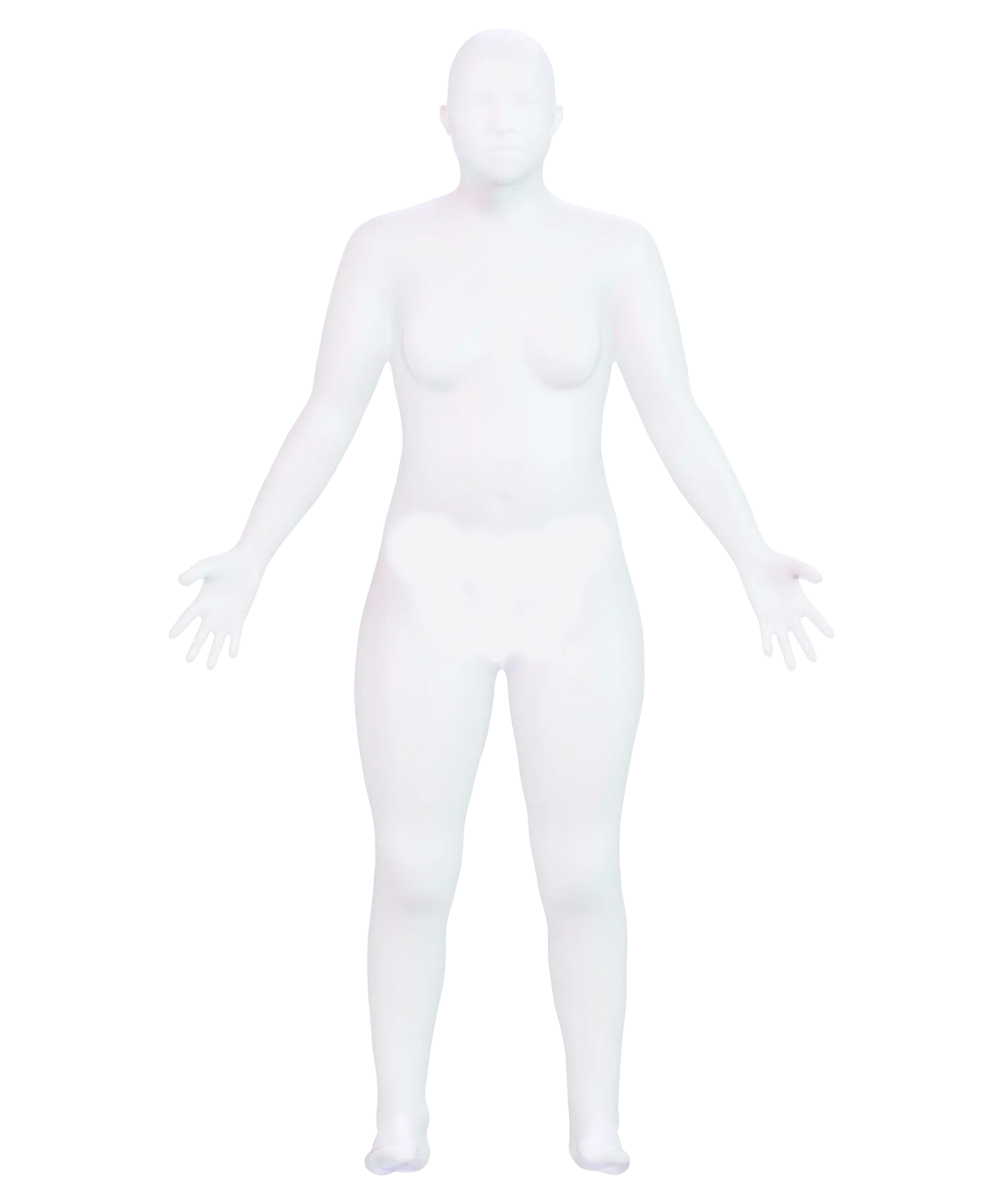
Rhabdomyolysis is the breakdown of muscle fibers. As the fibers break apart, they release myoglobin, a muscle protein, into the blood. Follow it from the thigh, marked in red, to the kidneys, marked in blue.

Start the journey Scroll to zoom in

Body → muscle

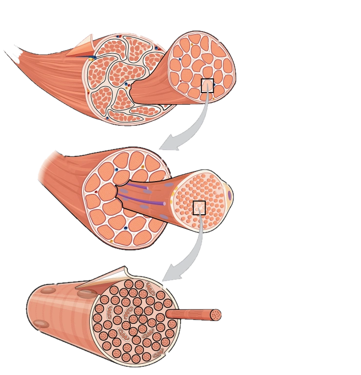
A muscle is made of bundles, and each bundle is made of fibers.

At the top, a muscle cut open shows how it’s divided into fascicles, bundles wrapped in connective tissue. In the middle, an opened fascicle reveals its fibers. At the bottom is a single muscle fiber, a long, cylindrical cell.

Muscle → fiber

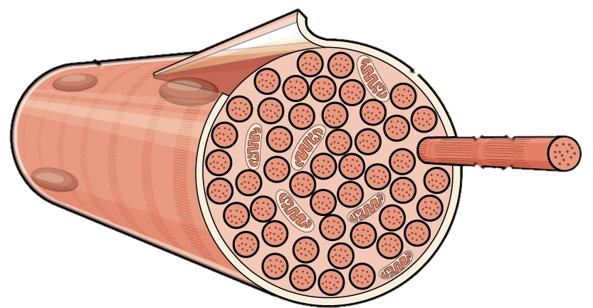
Each fiber keeps its contents behind a membrane.

That membrane is called the sarcolemma. Inside the fiber is myoglobin, a protein that stores oxygen so the muscle can keep working. If the sarcolemma tears, myoglobin and other substances inside the fiber leak into the blood.

Blood → kidney

Myoglobin travels all the way to the kidneys.

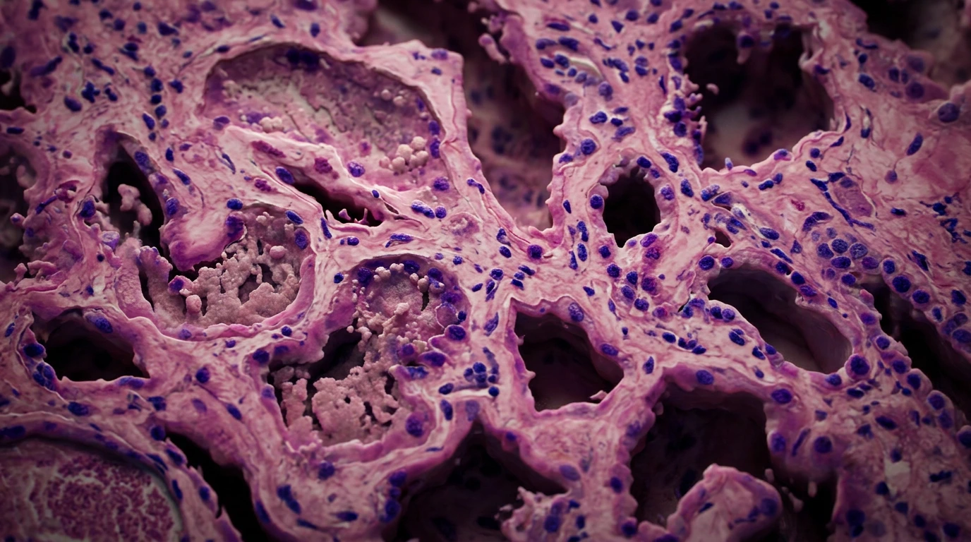
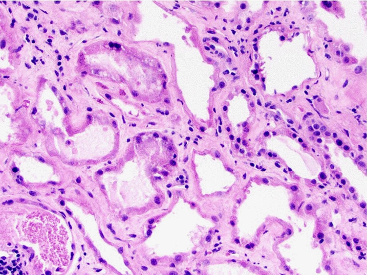
This is what a kidney affected by rhabdomyolysis looks like under the microscope: damaged tubules with material built up inside them. Below, you’ll see step by step how myoglobin gets there.

Follow the chain of events

The journey marks a thigh muscle and both kidneys on the body; shows how a muscle divides into fascicles and fibers; zooms in on one fiber to point out its membrane, the sarcolemma; and ends with damaged kidney tubules seen under the microscope.

How it happens, step by step

From muscle to kidney in three steps.

When many fibers break down, a lot of myoglobin reaches the bloodstream. The kidneys filter it out, and inside their tubules it can form plugs and damage the cells that line them.

Kidney damage is a common complication of rhabdomyolysis, but it doesn’t happen to everyone.

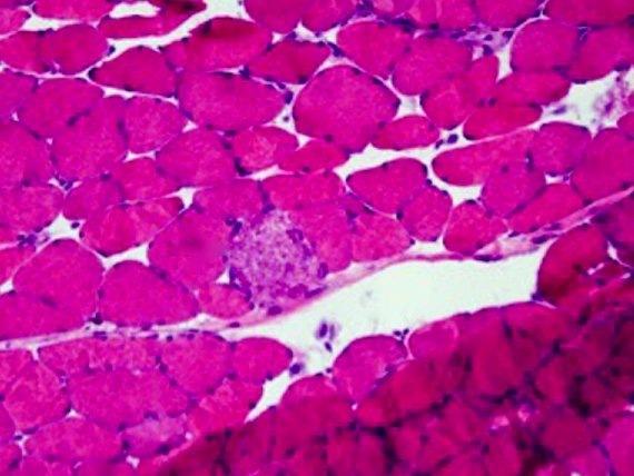
  1. Muscle biopsy stained with hematoxylin and eosin: pink, polygonal fibers and, in the center, a pale, granular fiber marked as necroticNecrotic fiberPale and granular: it has died

    A damaged fiber loses its shape.

    This biopsy comes from the thigh of a person whose rhabdomyolysis was caused by a statin, a cholesterol-lowering medicine. It’s stained with hematoxylin and eosin, the routine stain that colors muscle pink and nuclei purple. The surrounding fibers keep their polygonal shape. The marked fiber is pale and granular: it’s a necrotic fiber, a muscle cell that has died.

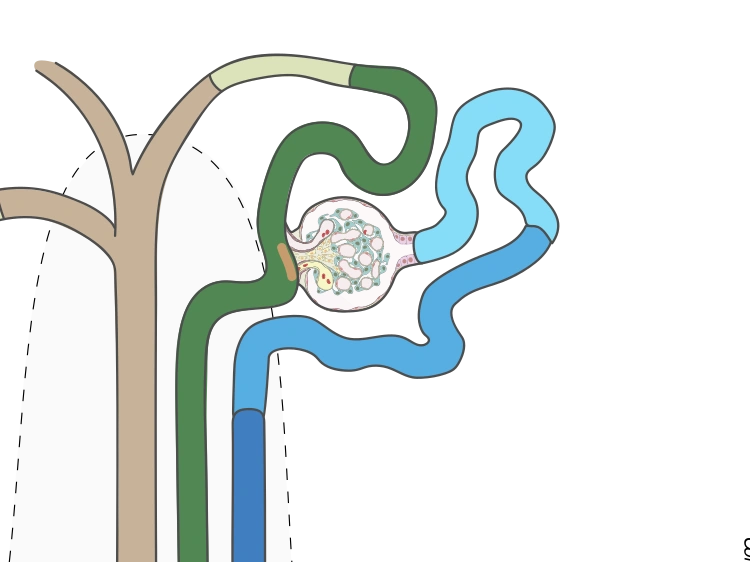
  2. Illustration of a nephron: the glomerulus, a round tuft of blood vessels, and the tubules that lead away from itGlomerulusEach nephron’s filter

    Blood carries myoglobin to the kidney’s filters.

    Each kidney contains many nephrons like the one in this drawing. Each nephron begins at a glomerulus, which filters the blood, and continues into a tubule that returns what the body needs to the bloodstream.

    Myoglobin is a small protein, so it passes through the glomerulus and into the tubule. Normally the tubule takes it back up, but when too much arrives, the tubule can’t keep up.

The third step happens inside the tubule. See it under the microscope.

Inside the tubule

Myoglobin leaves its mark in the tubule.

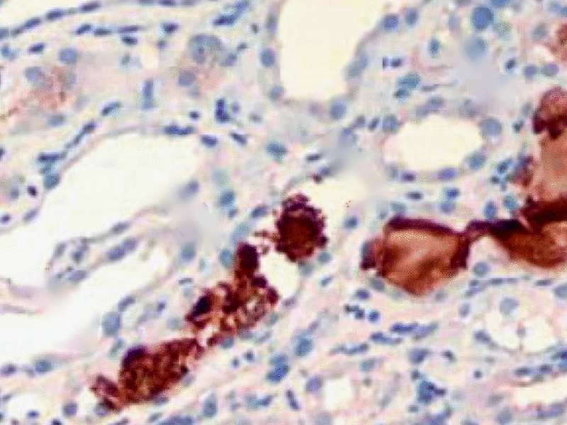
Switch between three microscope images: healthy tubules, damaged tubules, and the same damaged kidney treated with a stain that turns myoglobin brown.

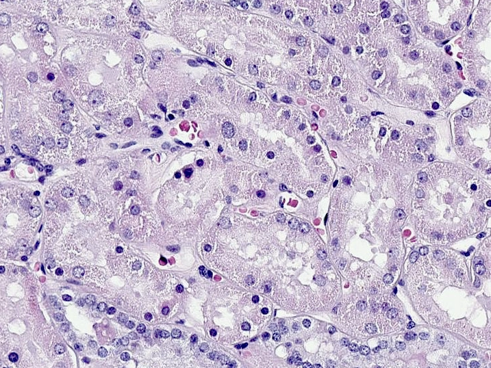
Normal human kidney cortex stained with hematoxylin and eosin: tubules lined by a thick layer of cells around a small central space
Healthy tubules: a thick ring of cells.

The cortex of a normal human kidney, stained with hematoxylin and eosin. Cut crosswise, each tubule is a ring of pink cells with purple nuclei surrounding a small central space called the lumen.

Kidney biopsy from a person with rhabdomyolysis, stained with hematoxylin and eosin: dilated, thin-walled tubules containing granular materialGranular castMaterial filling the tubule
Damaged tubules: thin walls and a filled lumen.

A kidney biopsy from a person with rhabdomyolysis. The tubules look dilated, and their cells are flattened. Inside is granular material: casts, which take the shape of the tubule as they form inside it and can block it.

Immunohistochemical stain for myoglobin on the same kidney biopsy: brown-stained casts inside the tubulesMyoglobin castThe stain turns it brown
The brown is myoglobin.

On the same biopsy, immunohistochemistry, a stain that uses antibodies, colors myoglobin brown. The casts filling the tubules stain brown, which shows that the material came from muscle.

Myoglobin binds to a protein made by the tubule itself, forming those casts. It also damages the tubule’s cells directly.

The damage starts in a muscle fiber. The evidence turns up in the kidney.

Key takeaways

Knowing the signs helps you act in time.

Some habits lower your risk, and recognizing rhabdomyolysis early means treatment can start sooner. It’s worth knowing this before it happens to you.

Get care right away

Just one of these three signs is reason enough.

Muscle pain or cramps far worse than you’d expect; dark urine the color of tea or cola, or urinating much less than usual; or weakness that keeps you from finishing activities you used to be able to do. These signs can appear hours or days after the muscle injury, and they can be mistaken for heat cramps or dehydration.

How it’s confirmed

With repeated blood tests. A urine dipstick isn’t enough.

Symptoms alone can’t confirm it. Doctors measure creatine kinase (CK), an enzyme that leaks out of damaged muscle, and repeat the test to see whether the level is rising or falling. A urine dipstick can’t rule out rhabdomyolysis: myoglobin is cleared quickly and may not show up.

Who’s at higher risk

Extreme exertion isn’t the only cause.

Intense exertion, such as running a marathon, can cause it. So can extreme heat, hard blows to the body, and certain drugs and medications, including statins.

Physically demanding work, some infections, and supplements such as creatine or ephedra also raise the risk.

Medical transparencySources, licenses, and scope

Every medical claim and every image in this journey links to a source you can open.

In the diagrams and micrographs, printed labels, arrows, and letters were removed with code, without redrawing any tissue. Brightness was matched between the normal and damaged kidney images, and the markers you see on this page were drawn on top.

The normal kidney image comes from the Human Protein Atlas, from a normal-looking area of a human surgical specimen. It serves as a reference for what healthy tissue looks like, not as a before-and-after comparison. The hematoxylin and eosin image of the injury and the myoglobin stain come from the same published kidney biopsy, from a case of rhabdomyolysis associated with COVID-19. The muscle biopsy comes from a different published case, in which a statin caused the rhabdomyolysis.

This journey explains how rhabdomyolysis develops and what its warning signs are. It doesn’t diagnose the condition, replace a medical evaluation, or recommend treatment for any individual.

Open-access sources, verified on October 2, 2026.

Know someone who would find this helpful?

Share it with them.