
Thyroid · Visual journey

It is rarely cancer. It still deserves a closer look.

From the neck to the different forms a nodule can take.

Begin the journeyScroll to change scale

Organ

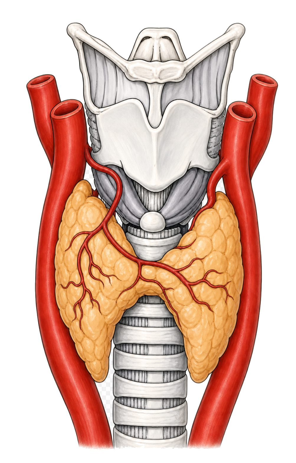
The thyroid.

It sits in the neck below the Adam’s apple, wrapping around the trachea. Two lobes are joined by a narrow band.

Tissue

The follicle.

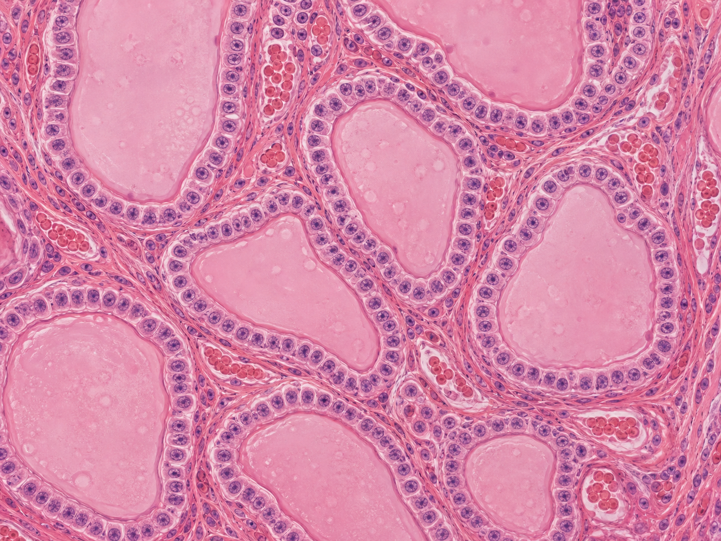
Inside are thousands of follicles. The pink material is colloid: it stores thyroglobulin, the raw material used to produce T3 and T4.

Under the microscope

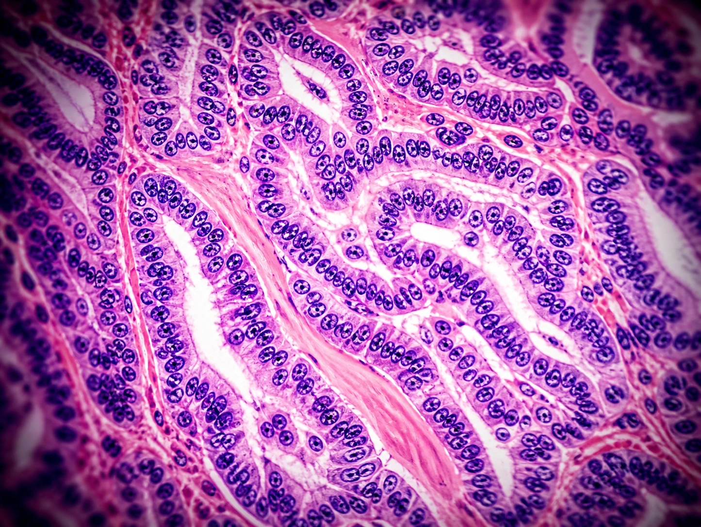
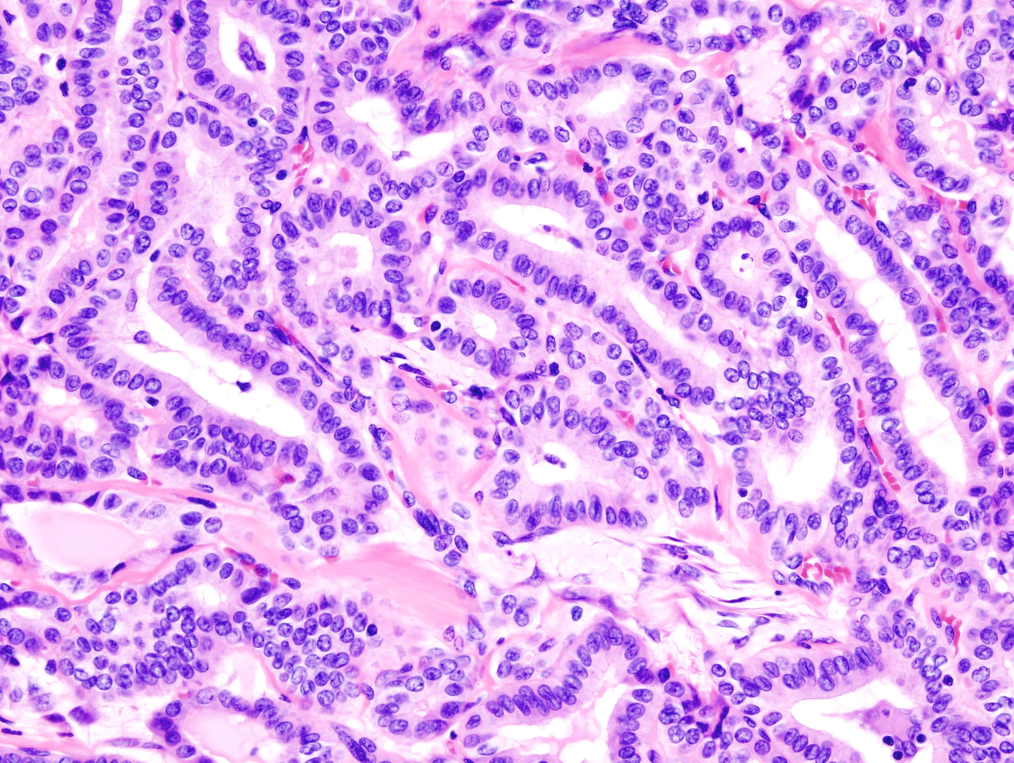
Papillary carcinoma.

In this lesion, cells form complex structures and their nuclei change. First we will see how it differs from normal thyroid and a benign nodule.

Compare the tissue

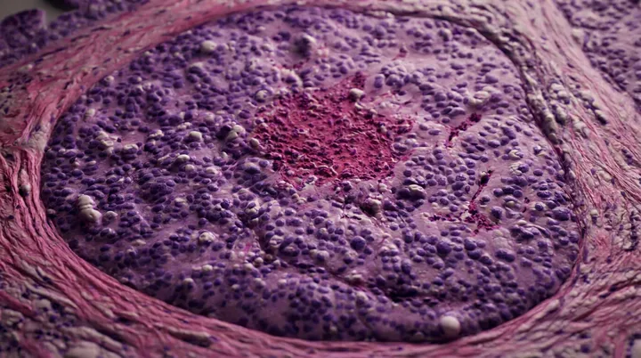
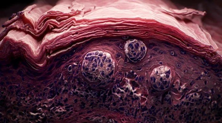
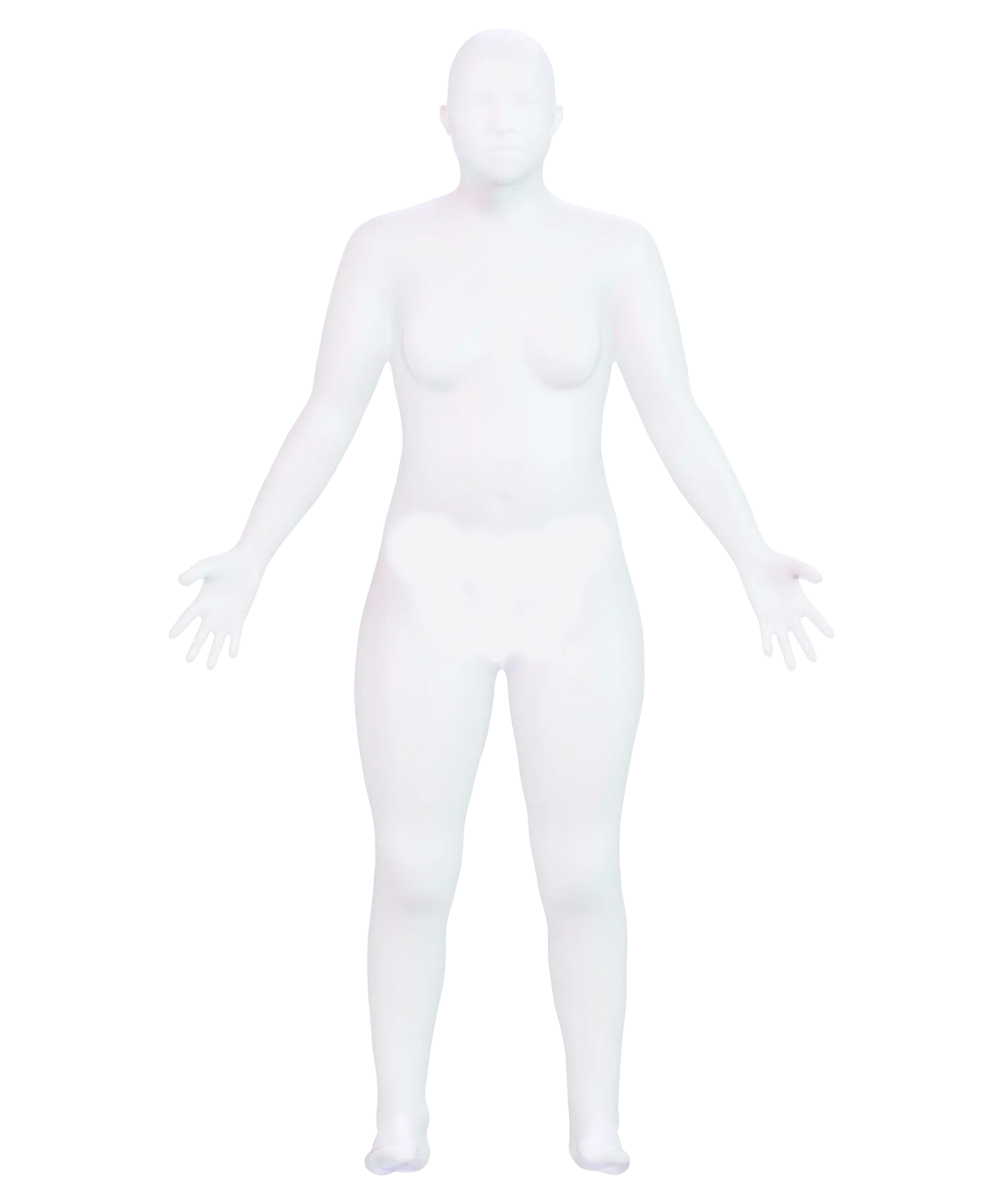
The sequence moves through the body, an illustration of the thyroid in the neck, a follicle with its ring of cells, and a real biopsy with papillary thyroid carcinoma.

Compare

The difference is in the architecture.

More than 90% of thyroid nodules are benign. We will first see normal thyroid, then a benign nodule, and finally papillary carcinoma, the most common thyroid cancer.

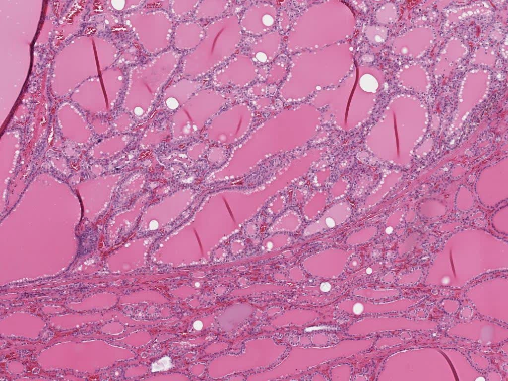
Thyroid micrograph with round follicles filled with colloidFairly uniform follicles
Unremarkable thyroid

The follicles contain pink colloid and retain a single layer of surrounding cells.

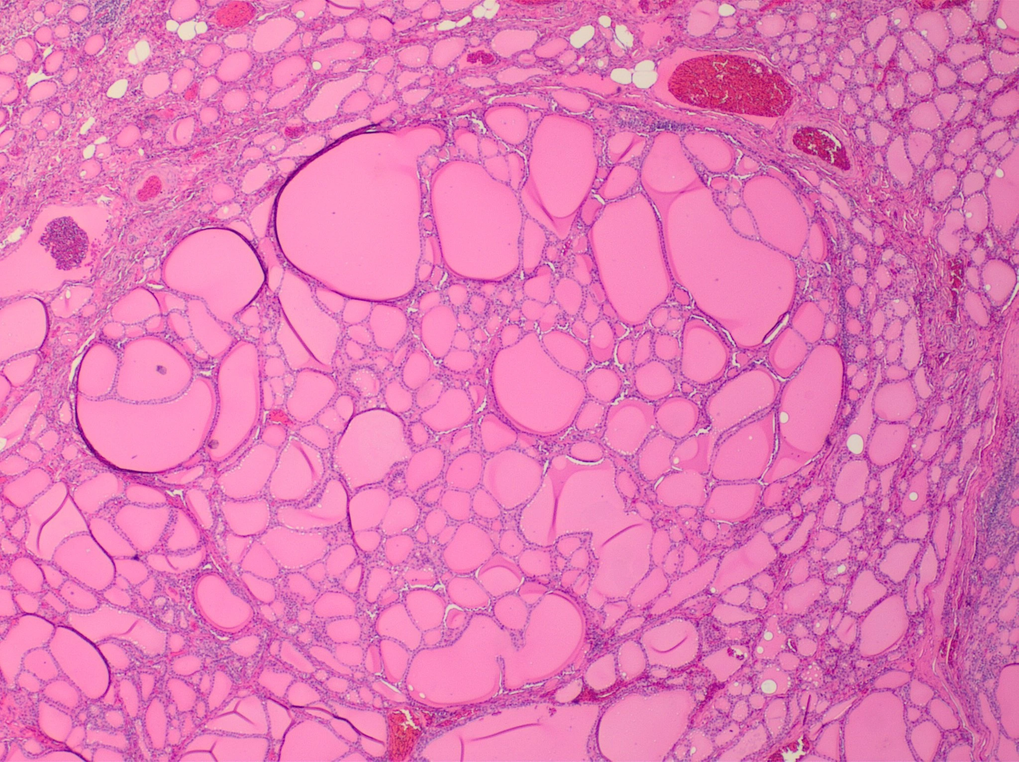
H&E micrograph of a benign hyperplastic thyroid nodule with follicles of varying sizesDifferent sizes
Benign hyperplastic nodule

The follicles are still present, but they grow unevenly. This is a common change and is not cancer.

H&E micrograph of papillary thyroid carcinoma with crowded cells lining folds of tissuePapillae and altered nuclei
Classic papillary carcinoma

It is the most common thyroid cancer. It forms folds that resemble small branches, and its nuclei change.

Know someone who would find this helpful?

Share it with them.