

Aging · Visual journey

What aging looks like.

No one shows you aging; they describe it with numbers. Here you will see it in five tissues.

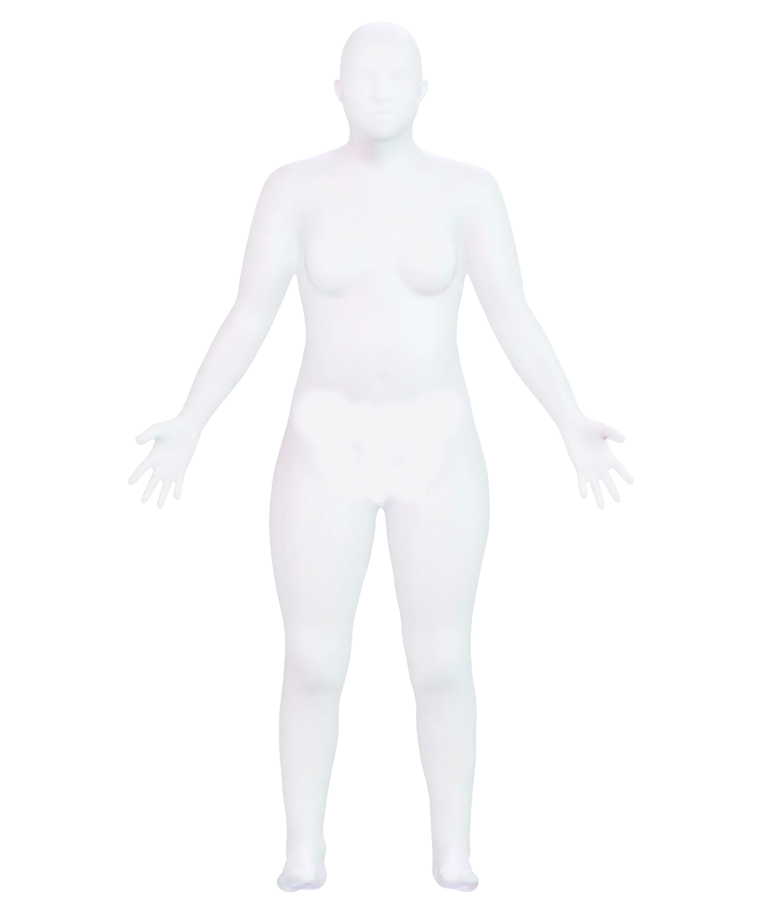
Three points are lit: the neck, chest, and thigh. We will examine an artery, the heart, muscle, bone, and the marrow inside it.

Begin the journeyScroll to change scale

The artery

A wall that expands.

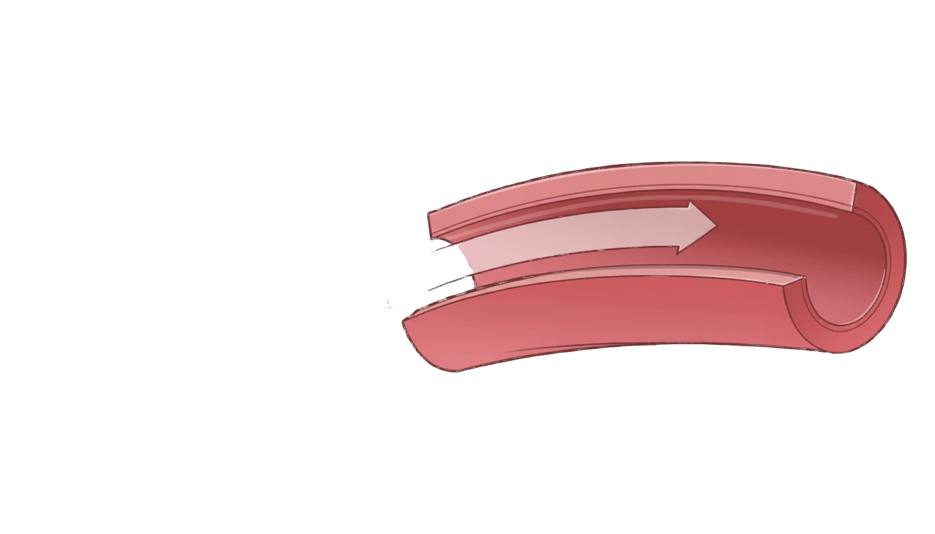
With each heartbeat, the arterial wall expands as it receives blood and then returns to shape. That elastic recoil cushions the pulse; over time, especially in large arteries, the wall can become stiffer.

The cell

Cells that last for decades.

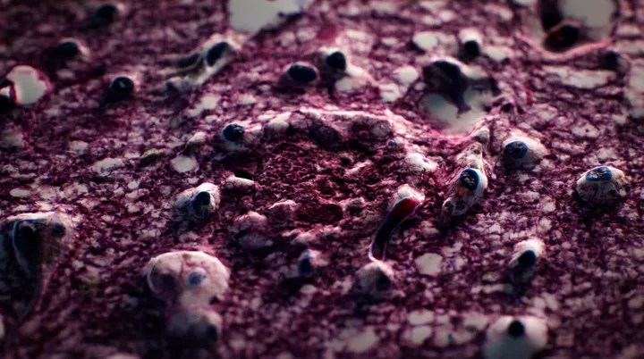
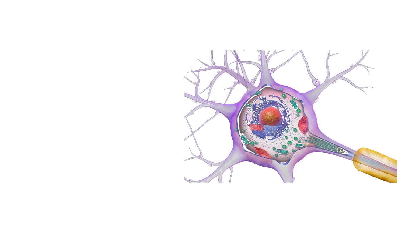
Neurons and cardiomyocytes, the muscle cells of the heart, are replaced very little. Their lysosomes contain enzymes that break down and recycle cell components; some material they cannot degrade can accumulate over time.

Under the microscope

Lipofuscin accumulates.

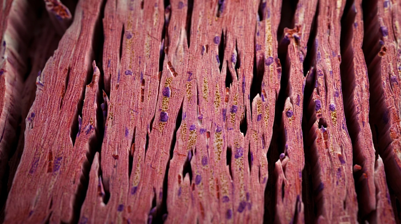
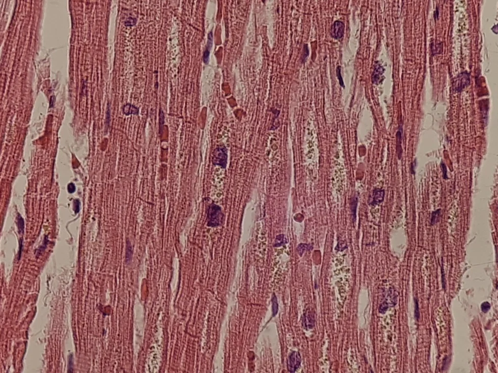
This is heart muscle. The yellow-brown granules next to the nuclei are lipofuscin: residual material that remains inside lysosomes after incomplete degradation. It accumulates especially in cells that divide very little, such as cardiomyocytes and neurons.

See the tissue comparisons

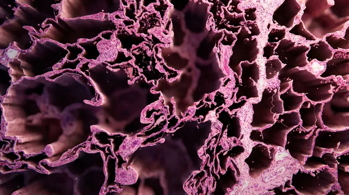
The sequence moves through the body with three marked points, a healthy artery opened from within, a neuron with its lysosomes, heart muscle with lipofuscin, and comparisons of trabecular bone and bone marrow.

What it looks like under the microscope

Five tissues, the same story.

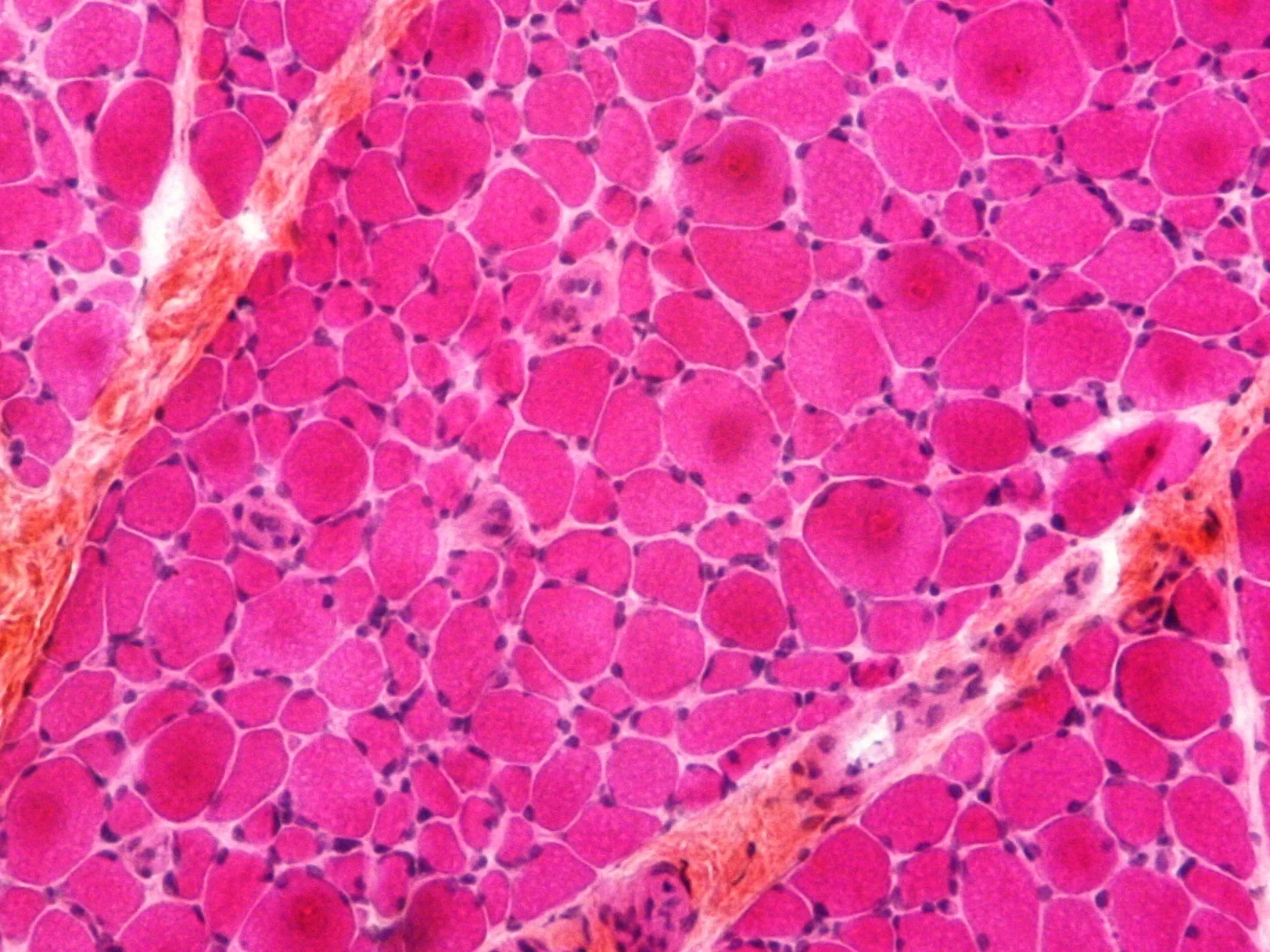
Four cards show hematoxylin and eosin (H&E), the most common stain in histology: eosin makes tissue pink and hematoxylin highlights nuclei in purple. The skeletal muscle slide uses HPS, a related stain with a more magenta appearance.

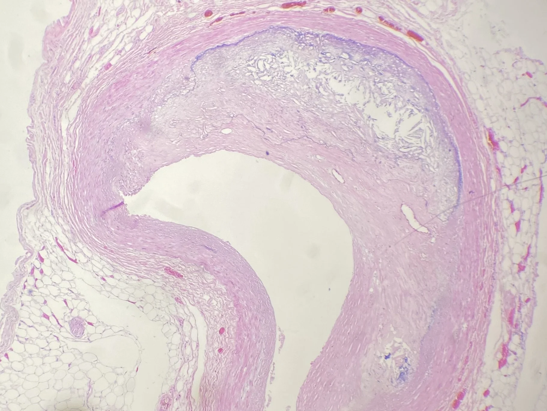
Micrograph of a coronary artery in cross-section with its wall thickened by plaqueThe artery
The wall has filled in

This is a coronary artery in cross-section. The atheromatous plaque is the pale, granular band that thickens the wall and narrows the space through which blood flows.

Atherosclerosis is the buildup of cholesterol and other material within the arterial wall. It becomes more common with age, but it is not a normal change caused simply by getting older.

Micrograph of muscle in cross-section, with large fibers beside groups of small angular fibersSkeletal muscle
Skeletal muscle atrophy

Each pink profile is a skeletal muscle fiber cut in cross-section. The small angular fibers are atrophic: they have lost volume.

With aging, muscle fibers can decrease in size and number, contributing to loss of mass and strength. This image shows how to recognize an atrophic fiber; by itself, it does not reveal the cause.

Micrograph of heart muscle with clusters of golden lipofuscin granules next to nucleiThe heart
Lipofuscin accumulates

These are heart muscle fibers cut longitudinally. The yellow-brown granules around some nuclei are lipofuscin, residual material that accumulates inside lysosomes when cellular degradation is incomplete.

Its amount often increases with age, especially in cells that rarely divide. It is a change associated with cellular aging; by itself, it does not mean heart disease is present.

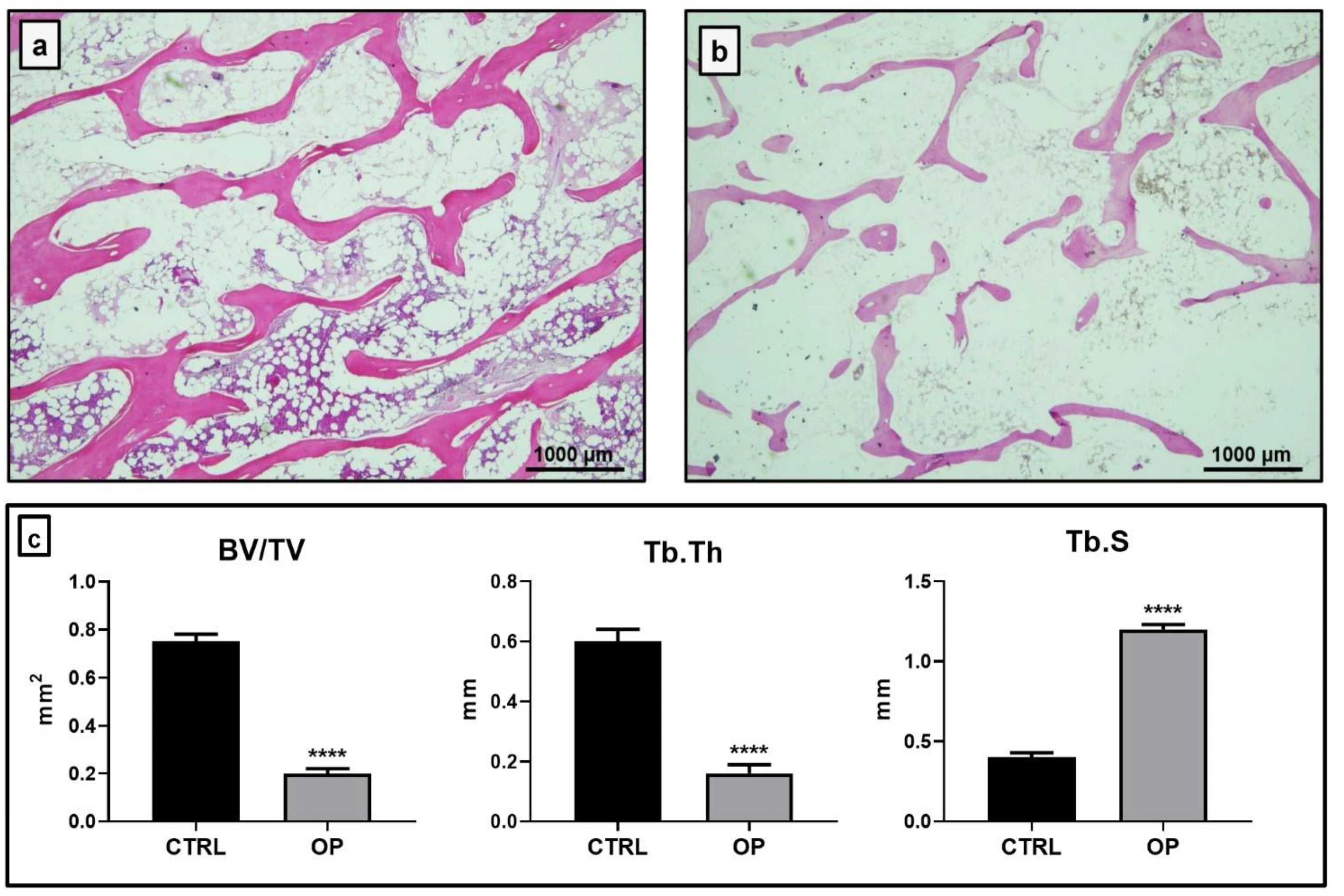
Human trabecular bone without osteoporosis, with thick connected trabeculaeBone without osteoporosisAbundant network
Human trabecular bone with osteoporosis, with thinner and more separated trabeculaeOsteoporosisThinned network
Osteoporosis leaves less internal network

Trabeculae are the pink bars that form the bone’s internal network. In bone without osteoporosis they are thicker and better connected; in osteoporosis they become thin, discontinuous, and farther apart.

Osteoporosis is a disease of low bone mass and deterioration of this architecture. It becomes more common with age, but it is not an inevitable consequence of aging.

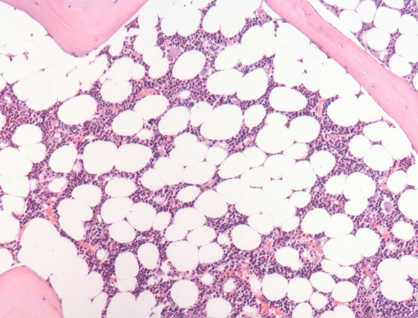
Human bone marrow with purple hematopoietic tissue between adipocytesHematopoietic tissueMakes blood cells
Human bone marrow rich in adipocytes, visible as large white spacesFat componentThe white spaces are adipocytes
Marrow combines forming blood cells and fat

The purple, highly cellular tissue is hematopoietic: blood cells form there. The large white spaces are adipocytes, cells that store fat; in this field they occupy about 60% of the marrow.

In people, bone marrow fat also increases with age. It shares the bone with blood-forming tissue and often increases as bone is lost, although neither change alone explains the other.

None of this hurts while it happens. That is why it helps to see it.

What you can influence

Not everything ages at the same pace.

These changes cannot be stopped completely, and anyone promising otherwise is selling you something. But they do not all carry the same weight: concrete actions can help protect arteries, muscles, and bones, while some cellular changes cannot currently be erased.

  1. 01The artery

    Over time, the aorta and other arteries become thicker, stiffer, and less flexible, which contributes to rising blood pressure. This hardening is called arteriosclerosis.

    • MoveStrength and aerobic exercise benefit the heart, and the most important thing is to move: any physical activity is better than none.
    • Do not smokeSmoking increases damage to arterial walls. Quitting, even later in life, can reduce risk.
    • Measure blood pressure and cholesterolBoth should be checked even when you feel well because they may cause no warning symptoms. Keeping them in range is among the most useful things you can do for your arteries.
  2. 02Muscle

    Lean tissue is lost over time: muscles lose cells. This loss is called atrophy.

    • Use resistanceStrengthening exercises can help maintain strength, slow muscle loss, and prevent falls.
    • Eat enoughWhen muscle shrinks from disuse, exercise and better nutrition can often reverse it. Age-related loss can be slowed, not undone; muscle needs enough material to rebuild.
  3. 03The cell that is not replaced

    Here we need to be direct. Cells that continue dividing distribute the pigment among daughter cells until it becomes negligible; neurons and heart muscle fibers rarely divide, so their pigment remains.

    • What does not existNo intervention has been shown to erase this pigment from a human cell that has already accumulated it: not supplements, diets, or therapies.
    • An honest clarificationAccumulation does not mean it is causing harm. It is currently read mainly as a mark of time; its role in cellular decline remains a hypothesis, and finding it in a biopsy does not by itself require additional testing.
    • What you can doProtecting circulation can benefit both the brain and heart, and controlling blood pressure may help delay or reduce dementia risk later in life. What reaches these cells can be influenced.
  4. 04Bone

    The trabecular network can thin and lose connections over time. This reduces bone strength, but the rate of loss is not the same for everyone.

    • Load the skeletonWalking, climbing stairs, and strengthening exercises make bone work and help preserve it.
    • Provide building materialBone needs enough calcium, vitamin D, and protein. Food is the first option; if a deficiency exists, a healthcare professional can advise how to correct it.
    • Prevent fracturesNot smoking, limiting alcohol, and preventing falls also protect what matters: preserving mobility and independence.
Medical transparencySources and scope

Medical evidence, visual provenance, and the editorial transformations applied.

The anatomical illustrations were cleaned to show one idea per frame: labels and guides were removed, and anatomy was not redrawn. The three original artery, muscle, and heart micrographs use only tighter framing. The bone comparison shows human sections identified by the source as control and osteoporosis. The marrow card enlarges two regions of the same human section to distinguish hematopoietic tissue from adipocytes; it does not present them as before and after.

Three clarifications about the slides. The muscle biopsy comes from a case in which fibers lost their nerve connection, and it uses HPS stain, which appears more magenta than routine H&E. It was chosen because the same field shows full-sized fibers beside shrunken fibers, a change that also occurs with aging. The artery collapsed during processing, so the shape of its central space does not measure narrowing during life. Aging pigment is shown in heart muscle because cardiomyocytes, like neurons, are rarely replaced; no equivalent openly licensed neuron slide is available.

The modified neuron illustration and muscle crop are shared under the same CC BY-SA license as their originals. None of the three sources records the donor’s age, and none includes a healthy field of the same tissue for direct comparison. The images show the changes, not how much any one person will experience them.

This journey explains changes that can occur in the body over time. It does not diagnose your body or replace a medical consultation. The slides shown come from other people’s tissues.

Sources verified August 23, 2026.

Know someone who would find this helpful?

Share it with them.